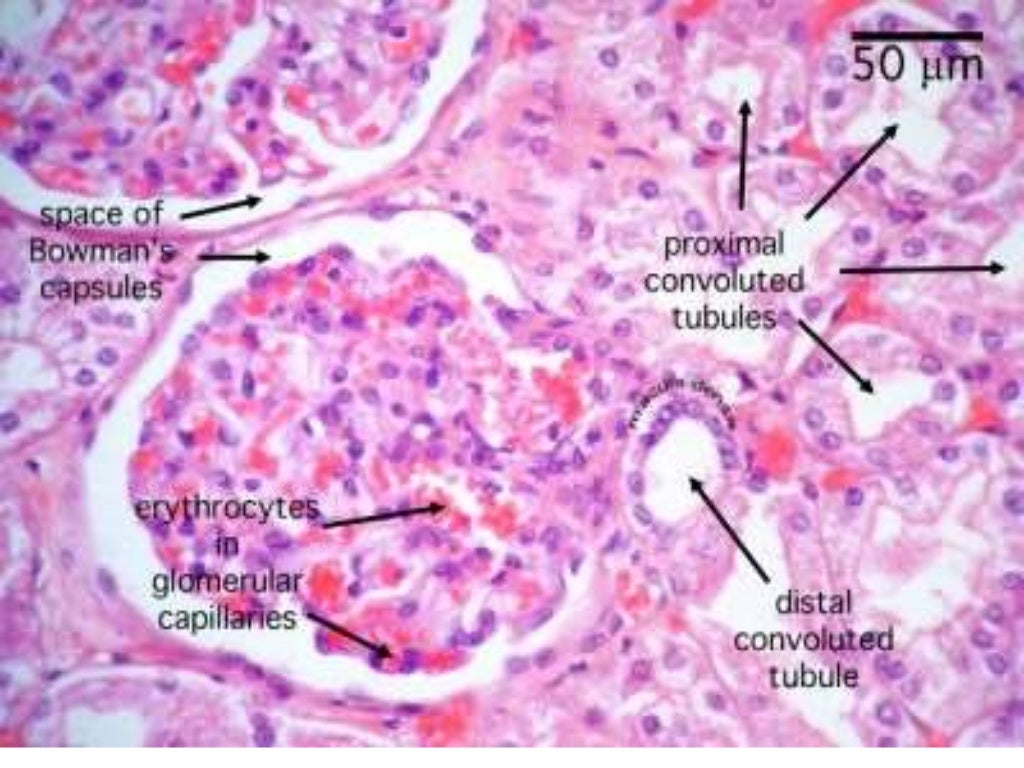
Kidney Microscope Slide Labeled. The renal structures that conduct the essential work of the kidney cannot be seen by the naked eye. describe the histology of the proximal convoluted tubule, nephron loop, distal convoluted tubule, and collecting duct. histology of connective tissue (capsule, cortex, medulla) in a kidney stained with azan. The outer region or renal cortex, containing many small, round renal corpuscles, much of the. the nephron is the main functional unit of the kidney, in charge of removing metabolic waste and excess water from the blood. Only a light or electron microscope can reveal these structures. Learning about kidney histology doesn’t have to be as painful as kidney stones! In this article we will explore the microanatomy of a nephron and learn how their function relates to their histological features. in this virtual slide of kidney you should be able to identify. microscopic structures mostly found in the renal cortex, but some components found in the medulla;
from www.slideshare.net
histology of connective tissue (capsule, cortex, medulla) in a kidney stained with azan. Only a light or electron microscope can reveal these structures. The outer region or renal cortex, containing many small, round renal corpuscles, much of the. microscopic structures mostly found in the renal cortex, but some components found in the medulla; the nephron is the main functional unit of the kidney, in charge of removing metabolic waste and excess water from the blood. in this virtual slide of kidney you should be able to identify. Learning about kidney histology doesn’t have to be as painful as kidney stones! describe the histology of the proximal convoluted tubule, nephron loop, distal convoluted tubule, and collecting duct. The renal structures that conduct the essential work of the kidney cannot be seen by the naked eye. In this article we will explore the microanatomy of a nephron and learn how their function relates to their histological features.
Kidney histology
Kidney Microscope Slide Labeled describe the histology of the proximal convoluted tubule, nephron loop, distal convoluted tubule, and collecting duct. in this virtual slide of kidney you should be able to identify. Only a light or electron microscope can reveal these structures. describe the histology of the proximal convoluted tubule, nephron loop, distal convoluted tubule, and collecting duct. microscopic structures mostly found in the renal cortex, but some components found in the medulla; In this article we will explore the microanatomy of a nephron and learn how their function relates to their histological features. histology of connective tissue (capsule, cortex, medulla) in a kidney stained with azan. the nephron is the main functional unit of the kidney, in charge of removing metabolic waste and excess water from the blood. The outer region or renal cortex, containing many small, round renal corpuscles, much of the. Learning about kidney histology doesn’t have to be as painful as kidney stones! The renal structures that conduct the essential work of the kidney cannot be seen by the naked eye.